Case Presentation
Clinical History
A 5-year-old female presented with a complaint of urinary retention.
Ultrasound examination revealed a large, well-defined heterogeneous pelvic lesion measuring 11.7 × 9.8 × 6.6 cm, demonstrating internal vascularity. The lesion was noted to be displacing both ovaries and the urinary bladder superiorly.
Serum Alpha-Fetoprotein (AFP) and Beta Human Chorionic Gonadotropin (β-HCG) levels were within normal limits.
Diagnostic Imaging
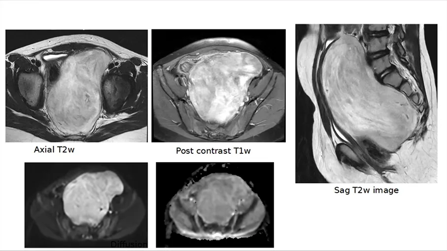
Diagnostic Challenge